
Page 111 - 广西植物2024年1期
P. 111
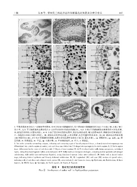
1 期                  朱永等: 紫纹兜兰的花形态和双雄蕊花药发育及分类学意义                                            1 0 7


             1. 早期花药原基分化出一对侧生并列药室ꎬ中央分化为不育隔膜组织ꎬ每个药室被不育隔膜组织分成 2 个小孢子囊ꎬ小孢子囊呈
             浅 U 型ꎮ 2ꎬ3. 不育隔膜组织边缘分化出 1~ 2 层具双核的内绒毡层细胞(3)ꎮ 4ꎬ5. 小孢子母细胞减数分裂期间的 4 层花药壁ꎬ
             注:绒毡层为双核(白箭头所指)ꎮ 6-9. 小孢子四分体时期的花药壁ꎬ包括齿状的表皮、增大的药室内壁、降解的中层和绒毡层ꎬ
             注: 绒毡层具双核(白箭头所指)ꎮ 10. 成熟花药时的花药壁ꎬ示表皮降解ꎬ药室内壁纤维状加厚ꎮ 11ꎬ12. 成熟花药的纵切面
             (11)和横切面(12)ꎬ示中央不育隔膜组织降解ꎬ花药室顶部花药壁变薄并开裂(11ꎬ箭头所指)ꎮ cn. 药隔组织ꎻ ep. 表皮ꎻ en. 药
             室内壁ꎻ it. 内绒毡层ꎻ m. 中层ꎻ sp. 小孢子囊ꎻ ss. 不育隔膜组织ꎻ st. 柱头ꎻ t. 绒毡层ꎮ
             1. Two anther primordia surrounding a stigmaꎬ indicating each containing a pair of lateral juxtaposed thecaeꎬ a densely ̄stained microsporangia was
             differentiated into a sterile septum at centralꎬ and each theca was divided into 2 U ̄shaped microporangia by the sterile septum. 2ꎬ 3. Sterile septum
             tissue differentiated in the center of each theca with 1-2 layers of inner tapetum (3). 4ꎬ 5. Developed anther walls during sporogenesisꎬ including 4
             layersꎬ noting binucleated tapetum (white arrow indicated). 6-9. Anther walls at microspores developedꎬ containing toothed epidermis and enlarged
             endotheciumꎬ while middle layer and tapetum degradedꎬ noting binucleated tapetum (white arrow indicated). 10. Anther wall at ripened anther
             stageꎬ indicating debris of epidermis and fibrously thickened endothecium. 11ꎬ 12. Longitudinal (11) and cross (12) sections of ripened antherꎬ
             indicating a slit of each theca and collapsed sterile septum (11ꎬ arrow indicated). cn. Connective tissueꎻ ep. Epidermisꎻ en. Endotheciumꎻ it. Inner
             tapetumꎻ m. Middle layerꎻ sp. Sporangiaꎻ ss. Sterile septumꎻ st. Stigmaꎻ t. Tapetum.
                                              图版 Ⅱ  紫纹兜兰的花药壁发育
                                   PlateⅡ  Development of anther wall in Paphiopedilum purpuratum