
Page 198 - 《广西植物》2024年第5期
P. 198
9 8 6 广 西 植 物 44 卷
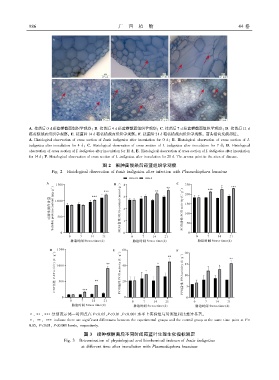
A. 接菌后 0 d 菘蓝横截面组织学观察ꎻ B. 接菌后 4 d 菘蓝横截面组织学观察ꎻ C. 接菌后 7 d 菘蓝横截面组织学观察ꎻ D. 接菌后 11 d
菘蓝横截面组织学观察ꎻ E. 接菌后 14 d 菘蓝横截面组织学观察ꎻ F. 接菌后 21 d 菘蓝横截面组织学观察ꎮ 箭头指向发病部位ꎮ
A. Histological observation of cross section of Isatis indigotica after inoculation for 0 dꎻ B. Histological observation of cross section of I.
indigotica after inoculation for 4 dꎻ C. Histological observation of cross section of I. indigotica after inoculation for 7 dꎻ D. Histological
observation of cross section of I. indigotica after inoculation for 11 dꎻ E. Histological observation of cross section of I. indigotica after inoculation
for 14 dꎻ F. Histological observation of cross section of I. indigotica after inoculation for 21 d. The arrows point to the sites of disease.
图 2 根肿菌侵染后菘蓝组织学观察
Fig. 2 Histological observation of Isatis indigotica after infection with Plasmodiophora brassicae
∗、∗∗、∗∗∗分别表示同一时间点在 P<0.05、P<0.01、P<0.001 水平上实验组与对照组相比差异显著ꎮ
∗ꎬ ∗∗ꎬ ∗∗∗ indicate there are significant differences between the experimental groups and the control group at the same time point at P<
0.05ꎬ P<0.01ꎬ P<0.001 levelsꎬ respectively.
图 3 接种根肿菌后不同时间菘蓝叶生理生化指标测定
Fig. 3 Determination of physiological and biochemical indexes of Isatis indigotica
at different time after inoculation with Plasmodiophora brassicae